Home

Nerf lent g onion skin microscope lentille téméraire Débutant

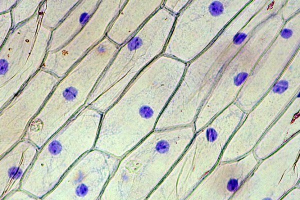
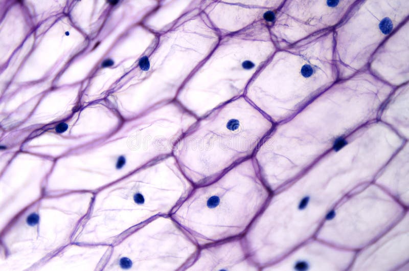
LM of Onion Skin - Stock Image - C012/1141 - Science Photo Library

OpenSciEd Virtual Microscope

Staining of Onion Cell Nuclei | Microbehunter Microscopy

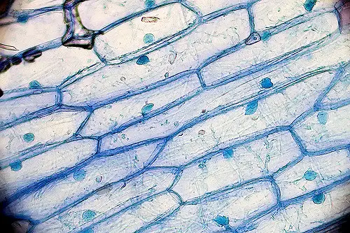
File:Onion skin cells under a microscope.jpg - Wikimedia Commons

Mic-UK: Homage to the onion skin

What difference will be observed under low and high powers of a microscope  during onion cell observation? - Quora
What difference will be observed under low and high powers of a microscope during onion cell observation? - Quora

School Science/How to prepare an onion cell slide - Wikibooks, open books  for an open world
School Science/How to prepare an onion cell slide - Wikibooks, open books for an open world

Onion Cells Under a Microscope - Requirements/Preparation/Observation

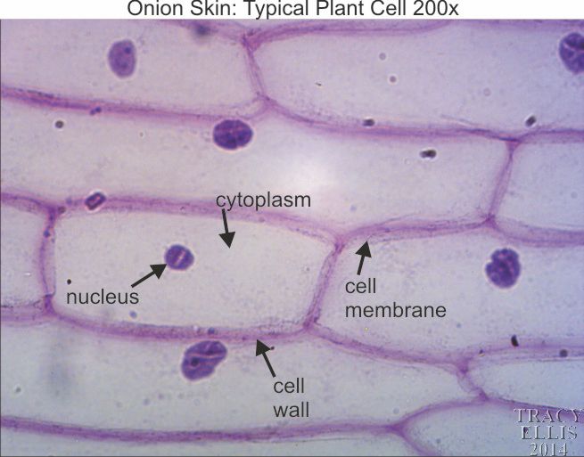
Onion skin 200x - Dissection Connection

Observing Onion Cells Under The Microscope » Microscope Club

Observing Onion Cells Under The Microscope » Microscope Club

Solved I did a bio lab where I looked at purple onion skin | Chegg.com

Onion Cells Under the Microscope - YouTube

Experiment on Onion Peel | Science Experiment | Conclusion As cell walls  and large vacuoles are clearly observed in all the cells, the cells placed  for observation are plant cells. | cell,
Experiment on Onion Peel | Science Experiment | Conclusion As cell walls and large vacuoles are clearly observed in all the cells, the cells placed for observation are plant cells. | cell,

1,350 Onion Microscope Images, Stock Photos, 3D objects, & Vectors |  Shutterstock
1,350 Onion Microscope Images, Stock Photos, 3D objects, & Vectors | Shutterstock

Onion skin showing cell during mitosis - Stock Image - B215/0032 - Science  Photo Library
Onion skin showing cell during mitosis - Stock Image - B215/0032 - Science Photo Library

351 Onion Skin Cell Images, Stock Photos, 3D objects, & Vectors |  Shutterstock
351 Onion Skin Cell Images, Stock Photos, 3D objects, & Vectors | Shutterstock

Onion Cell Microscope Slide Experiment - YouTube

Light Microscope Onion Cells Stock Photos - Free & Royalty-Free Stock  Photos from Dreamstime
Light Microscope Onion Cells Stock Photos - Free & Royalty-Free Stock Photos from Dreamstime

OpenSciEd Virtual Microscope

Onion Cells under Microscope

5. Compare the image of the unstained and stained onion skin cell under the  microscope.6. How did the iodine - Brainly.ph
5. Compare the image of the unstained and stained onion skin cell under the microscope.6. How did the iodine - Brainly.ph

Explore Scientific Smart Microscope Slide: Onion Bulb Epidermis (Engli

Salt Water and The Red Onion Root Lab | bulb